


The structure and functions of the organism
Skin
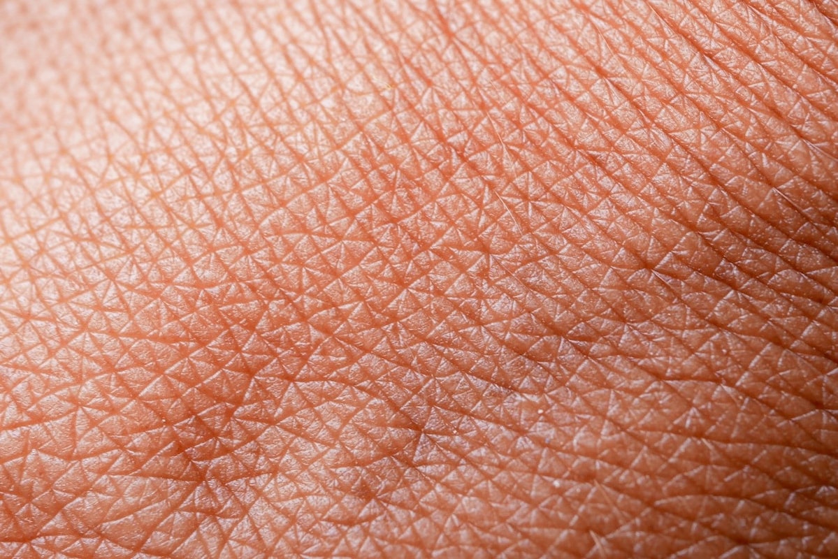
The skin protects the body from mechanical damage, from the effects of heat, cold and the sun, chemical effects and penetration of microorganisms. It also participates in the regulation of temperature - sweat cools the body and secretes harmful substances.
The skin is a dynamic, constantly changing organ that consists of three main layers: the epidermis, dermis, and subcutis, each composed of several sublayers. Skin extensions like the follicles and glands of the sweat and sebaceous glands also play a variety of roles in its overall function.
The composition of the skin includes nails and hair - at the body openings it transitions into a mucous membrane.
When examining a casualty, care should be taken to the continuity of the skin, i.e. the injury, the appearance of a rash and its colour, humidity and heat as indicators of the general condition of the organism.
Bones
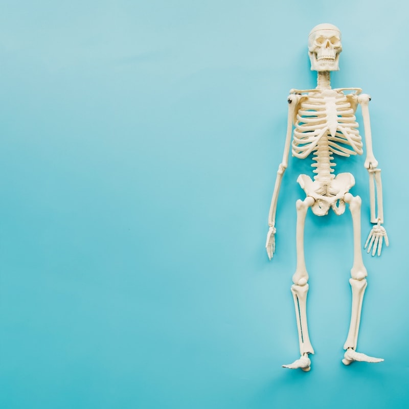
The skeleton serves as a support for the whole body, allowing movement and work**.**
The bones are composed of bone tissue that is hard on the surface and hollow as a sponge on the inside. the surface of the bone is surrounded by periosteum with nerve endings. In the cavity of the bone is a soft mass - bone marrow. The bone marrow of some bones produces blood elements.
Bones are connected to each other by joints, thus forming a skeleton that we can divide into the bones of the head, trunk and limbs.
The bones of the head include the bones of the skull and face. Between the bones of the head are mucous-lined cavities called sinuses.
The bones of the trunk consist of the spine, the pectoral bone of the ribs. The spine is made up of 33 vertebrae. The ribs are almost semi-circular bones - 12 pairs. They are attached to the spine with posterior joints and with cartilage to the chest.
The bones of the limbs are divided into the bones of the upper and lower limbs.
When examining casualties, we look for pain, swelling, deformity or some other alterations to the bone.
Joints
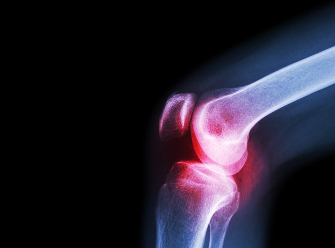
They allow the bone to move for movement, exercise, breathing and other functions. Joints are the head and cup of bone that fit into each other, coated with cartilage. They consist of articular cocoon, articular fluid and ligaments.
When examining casualties, we look for pain, deformity and swelling in the joints.
Muscles
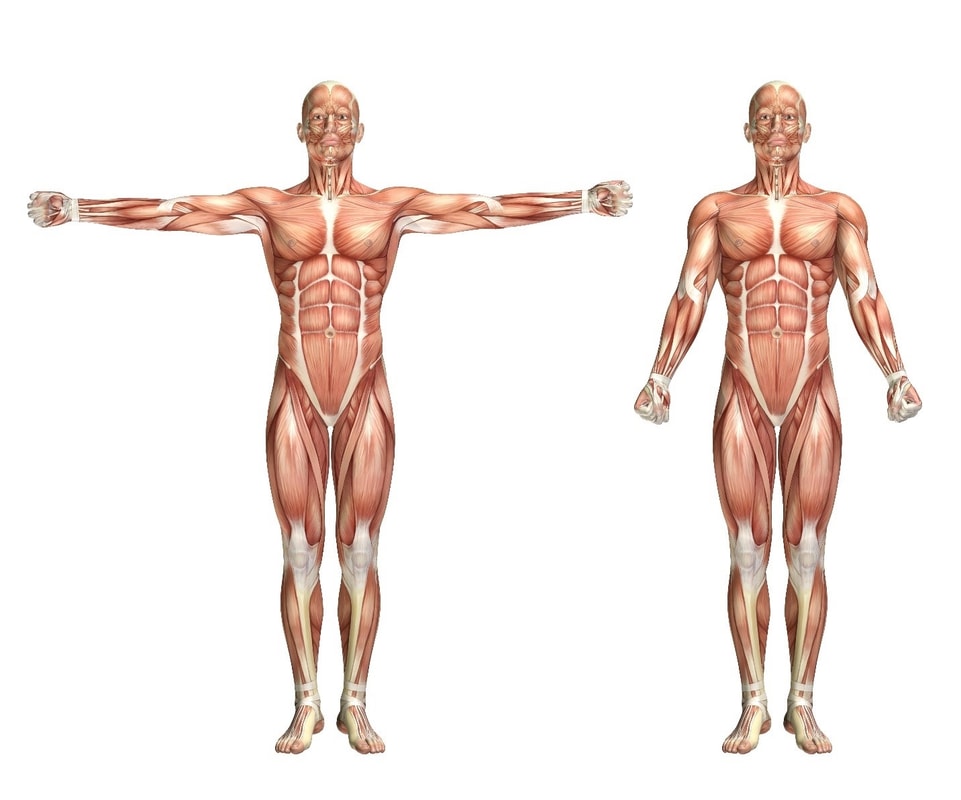
They are divided into three groups: transverse striped, smooth and cardiac. The transverse stripes are triggered willingly and are composed of smaller muscle fibers, which in turn are composed of yet smaller units called myofibrils. At their ends, the muscles turn into firm tendons, which attach them to the bones.
Smooth muscles are found in the internal organs: digestive, respiratory, blood vessels and many other. They set in motion all actions of the body that do not depend on our will.
The heart muscle is specially built and adapted to the function of the heart. It is made up of unique tissue that moves the heart.
In cases of muscle injuries, we also look for pain, deformity and swelling in casualties.
Respiratory system
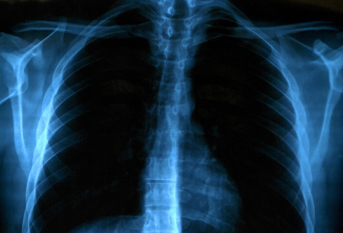
Respiratory organs include the nose, larynx, windpipe and trachea. The windpipe branches into two smaller airways, and these into many smaller twigs, which end in lungs.
Through the airways, air flows from the nose, through the trachea, smaller windpipes and the smallest branches all the way to the lung bubbles (alveoli). Lungs are surrounded by a network of thin arterial and venous blood vessels (capillaries).
Breathing is performed rhythmically by a muscular-membranous plaque - the ossicles or diaphragms and ribs driven by the rib muscles. The normal rhythm of breathing at rest is about 16 times per minute.
When inhaled, oxygen-rich air travels through the airways to the lungs. The lung bubbles deliver oxygen to the red blood cells (erythrocytes), and they distribute it throughout the body and deliver it to the cells where they are "burnt" to release energy. In this process, carbon dioxide (CO2) is produced, which is returned to the veins by blood and released into the environment by exhalation in the lungs.
The sense of smell is located in the nose, and should not be damaged in seafarers, because, among other things, it has the function of warning of danger.
Breathing emergencies can be caused by choking, a punctured lung, an allergic reaction, exposure to chemicals or other toxins, asthma, and other causes.
In casualties, we look for the existence of breathing, the rhythm of breathing, the existence of obstructions in the airways, the secretion and bleeding from the airways, the use of respiratory muscles, etc.
Cardiovascular system
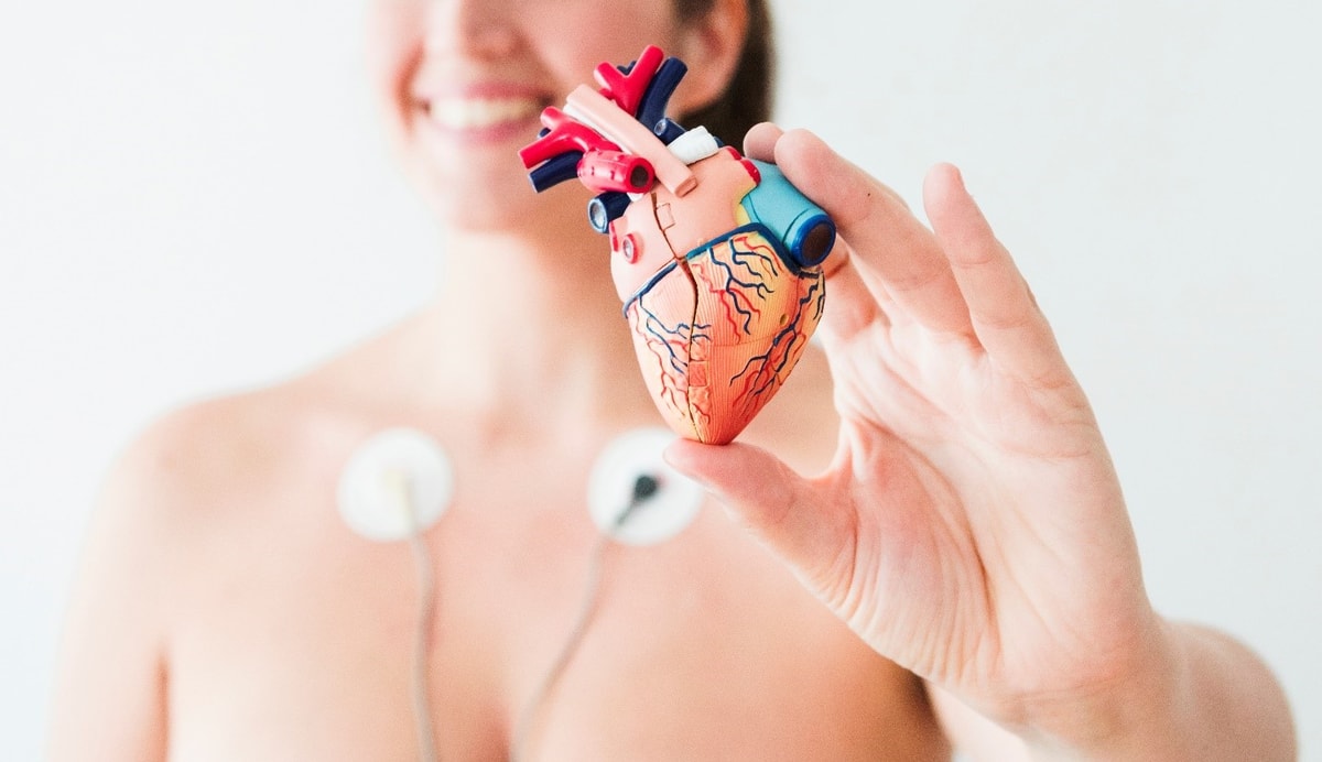
The circulatory system consists of three independent systems that work together: the heart (cardiovascular), lungs (pulmonary), and arteries, veins, coronary and portal vessels (systemic).
The heart is the "pump" that drives the blood through the whole organism. It is located in the thoracic cavity, behind the thorax with a slight shift to the left. It consists of the left and right atrium and left and right ventricle. The ventricles are larger than the atriums. There are cardiac valves between the unilateral pre-cell and the peritoneum whose function is to prevent blood from returning.
The pulse, which is usually felt on the arm, reflects the constriction of the left ventricle when blood from it presses into the arterial system. The pulse reflects the heartbeat, which normally beats 60 - 80 times per minute during rest.
Arteries are blood vessels that stream blood from the heart to the periphery of the body all the way to the cells themselves. They end with arterial capillaries. Arterial blood is light red in colour because it is rich in oxygen. Blood from the injured artery sprays in a jet.
The veins stream blood from the periphery to the heart. The venous system begins with the venous capillaries where the arterial capillary network ends. The veins merge into larger branches, and all the venous blood is collected in two large veins. The upper hollow vein collects blood from the upper parts of the body, and the lower hollow vein from the lower parts of the body.
An adult healthy person has 5-6 litres of blood in his body - it is composed of blood fluid (plasma) and blood cells - blood cells are divided into red (erythrocytes), white (leukocytes) and blood platelets (platelets).
Losing an excessive amount of blood is known as haemorrhagic shock. Doctors categorize haemorrhagic shock into four classes based on how much blood is lost. If a person loses more than 40 percent of his blood, he dies. This is about 2,000 mL, or 0.53 gallons of blood in the average adult.
Common problems of the circulatory system include an aneurysm, atherosclerosis, heart disease, high blood pressure and varicose veins.
A heart attack occurs when the blood flow that carries oxygen to the heart is blocked. The heart muscle becomes starved for oxygen and begins to die. Many heart attack patients die before they reach a hospital. Prompt medical treatment reduces the amount of heart damage.
Digestive system
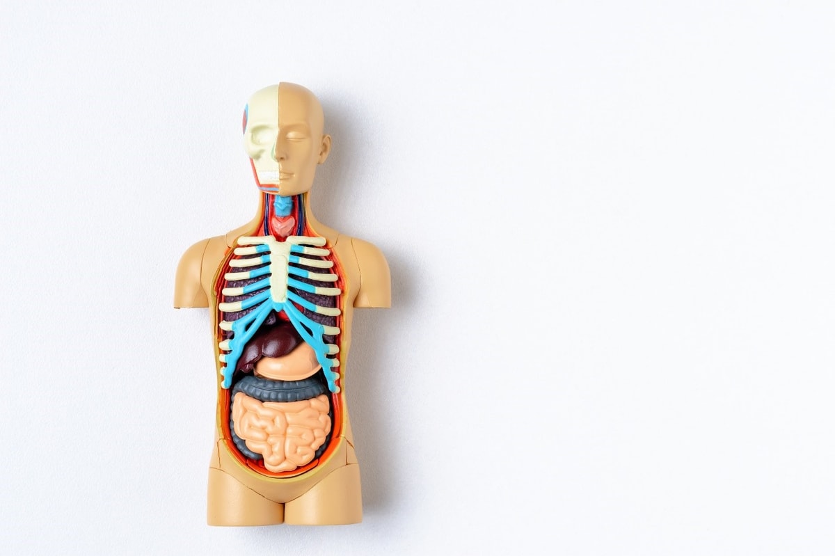
The digestive system begins with the oral cavity (teeth, tongue, salivary glands), followed by the pharynx, oesophagus, stomach, duodenum, small and large intestine. The small intestine is about 7 meters long. The colon is approximately 1.5 m long.
The digestive organs also include the liver and pancreas. The exit duct, through which the juices and pancreas are infused, is located in the duodenum.
In the oral cavity, the food is chewed and soaked in saliva, in the stomach it is digested by gastric juices, in the duodenum it additionally digested by the liver and pancreas juices, then in the small intestine the nutrients (intestinal fibrils) are absorbed, and in the large intestine water is absorbed and stool is formed (from indigested residues).
Certain gastrointestinal disorders can be life threatening and require emergency treatment. For many people, emergency treatment involves surgery.
Gastrointestinal emergencies are usually accompanied by severe pain. If a person has abdominal pain, he should be taken to a doctor who will decide what further steps to take. Emergency surgery of the abdomen is usually done when the abdominal pain seems to result from:
- An intestinal obstruction
- A ruptured or perforated (punctured) organ, such as the stomach, appendix or intestine
- A hernia with too little blood flow
- A blockage of blood flow
- An abdominal abscess (a pus-filled pocket of infection)
If you suspect a pain resulting from appendicitis or any of the above, seek immediate medical attention.
Urinary system
It consists of two kidneys and a ureter, a bladder and an urethra. The urinary tube is very short (about 3 cm) in women and longer in men. In men, the urethra passes through the prostate gland or the prenatal gland, whose function is to participate in the formation of seminal fluid.
A large amount of blood passes through the kidneys, which is supplied by the renal arteries and drained by the renal veins. The kidneys serve as a filter, eliminating the harmful products of metabolism. The kidneys remove the harmful components from the urine, which passes through the ureter and collects in the bladder. A healthy adult 4 to 6 pounds a day urinates 1 to 2 litres of urine.
Emergencies of the urinary tract centre on three major issues, including uncontrolled renal haemorrhage, accumulation of urine within the peritoneal cavity or retroperitoneal space, and obstruction to urine outflow.
Nervous system
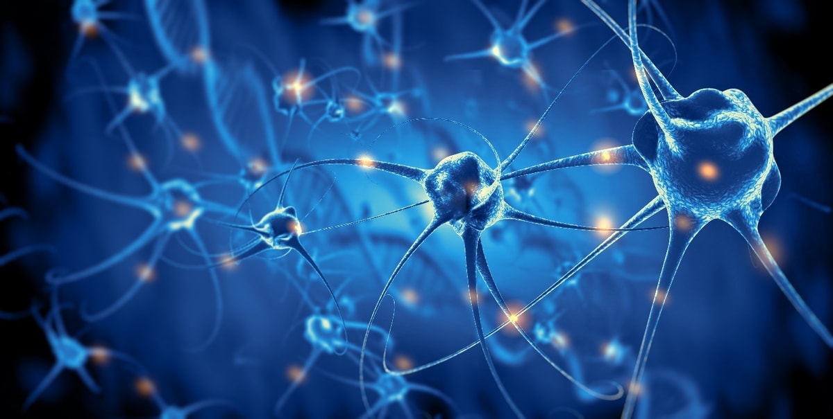
A nerve cell (neuron) is a basic element of the nervous system. Unlike other cells, nerve cells have many stellate branches and long extensions (nerve fibers) that contract into bundles and form nerves.
The nervous system is divided into central and peripheral. The central consists of the brain and spinal cord. The central nervous system is enveloped in brain sheaths and protected by bone armour - the skull and spinal column. A special fluid (CSF) circulates through the central nervous system. The brain is the centre of our thoughts, the interpreter of our external environment, and the origin of control over body movement.
The peripheral nervous system includes all the nerves in the body that lie outside of the spinal cord and the brain. These nerves carry information to and from the central nervous system to provide complex body functions. Sensory cells are involved in taking information from the periphery to the central nervous system.
The involuntary or autonomic nervous system is made up of nerves that move individual organs past our will. The autonomic nervous system drives the organs and systems of the respiratory system, digestion, heart, bloodstream and all other internal organs and glands.
The willing peripheral nerves have input fibres that receive stimuli from the outside world and transmit them to brain centres, from where the motor impulse emits a pulse that drives the muscle and performs the appropriate action.
There are many different causes, mild or severe, that can lead to a nervous system emergency. Some of these conditions and injuries include:
- Degenerative diseases (Parkinson's, Alzheimer's, MS)
- Infections
- Mental health problems (depression, anxiety disorders)
- Organ failure (e.g. heart failure)
- Problems with blood supply
- Trauma to the head or spinal cord
- Tumours
Common emergency examples are heart attack, seizure, stroke and spinal/head injuries.
Senses

The five senses usually attributed to humans are vision, hearing, taste, smell and touch.
The organs for the sense of sight are the eyes. They are made of eyeballs, eye muscles (move the eyeball in the desired direction) and eyelids (protective role). The image of the object being viewed is projected through the lens and intraocular fluid onto the retina, which sends the image through the optic nerve to the centre of vision in the brain (the eyes sees, the brain perceives).
The ears are the organ of hearing and balance, and consist of the outer, inner and middle ear. The auricle (pinna) is the visible portion of the outer ear which collects sound waves and channels them into the ear canal (external auditory meatus), where the sound is amplified. The sound waves then travel toward the eardrum, or tympanic membrane. Sound waves cause the eardrum to vibrate which sets the ossicles into motion - the three bones are named after their shapes: the malleus (hammer), incus (anvil) and stapes (stirrup). The sound waves enter the inner ear and then into the cochlea, a snail-shaped organ (inner ear) where nerve endings transform the vibrations into electrical impulses that then travel along the eighth cranial nerve (auditory nerve) to the brain. The inner ear also contains the vestibular organ that is responsible for balance.
The cells of the nasal mucosa make sense of smell. The sense of smell serves as a kind of chemical analyser that protects the body from spoiled food and other harmful gases and vapours.
The sense of taste is located in the tongue, whose surface is made up of about ten thousand taste buds, found on the papillae (the tiny bumps on the tongue).
The sense of touch and warmth is placed in the skin, especially at the fingertips.